As mentioned in the previous article, there are more than a million different types of microorganisms spread across different categories in the world. It is really essential to understand how to distinguish one from another; how to distinguish between a friend and a foe. The first step in understanding whether a particular species is harmful or not comes by preparing the specimen and analyzing it under the microscope. One cannot observe the sample under the microscope and get to know which is which. For this drawback, every specimen is well prepared using various steps to ensure that everything is detailed.
There are only a few steps to be followed in the preparation of a specimen out of which fixation and staining are the primary steps.
Fixation:
The stained cells or sample that is to be observed must resemble the living cells to an higher extent. Fixation helps in this. It is a process that inactivates enzymes to fix the ultrastructure internally as well as externally. The process disrupts the cell morphology and hardens the specimen making it clearly visible for the observer under microscope upon staining.
There are two types of fixation techniques. Heat fixation and chemical fixation.
1. Heat fixation: This technique is generally done for prokaryotes(a type of cell lacking nuclear membrane). Smear(a thin layer of cells) is taken onto a clean slide and is passed through the flame. This technique ensures the morphology is fixed but not the internal structures.
2. Chemical fixation: This technique is used to fix the substructures or structures inside the cell. The fixative penetrates through the membrane of the cell and chemically inerts the functioning of cellular components. Some common fixatives are ethanol, formaldehyde, etc.
Staining:
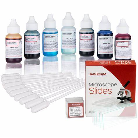
In simple terms, staining is a process used to color the components based on their chemical reactivity and visibility. While some structures can be directly seen without staining, some need either acidic or basic stains. Few specimens require negative staining; a case in which the background is stained making the specimen look brighter.
Staining is a simple task yet something that needs attention. There are different types of staining
1. Simple staining: It is the simplest type of staining. The required stain based on the chemical nature of the specimen is applied upon the smear and observed under the microscope by removing excess stain and covering the smear with a coverslip. Basic dyes are used in most cases when it comes to simple staining.
2. Differential staining: A type of staining used to distinguish between two closely related organisms. Classic examples are Gram staining and Acid-fast staining. The process includes the usage of two different stains that determines the type of organism based on the stain holding or cell composition of the organism. This includes a set of procedures that includes usage of mordants; a stain enhancer.
3. Structure specific staining: Some structures sometimes need special staining for clear visibility. Structures like capsules and endospores are hard to take stain as in other regular cases. Flagella needs to be stained specifically as it is one of the most delicate components and very minute to handle.
These steps ensure that the specimen is well prepared for observation under microscopes. The stain and fixation are to be done accurately to get the best possible vision under the microscope. If done, the results under the microscope will give a clear picture in the understanding of the functioning of or class of a specific organism.


0 Comments